CELL DYN 3500 is an electronic counter to measure the size,
shape, color, maturity, and other parameters with a very small amount of
blood in a few seconds i.e., the amount of blood required is less and the
result is obtained much faster compared to the manual method. The human error
factor is also less. The machine error factor is almost nil.
TECHNOLOGY USED:
-
Optical flow channel
-
Electrical impedence channel
-
Spectrophotometer
CELL DYN 3500 MEASURES
SAMPLE COLLECTION
2 ml blood in EDTA/Citrate bottle is to be sent to the Lab.
Do not inject the collected blood through the needle into the bottle. It may
damage the cells and results may become false.
Amount of blood needed by the machine is only 53 µL. So,
in a new born a smaller amount like 0.5 ml or 1 ml may sufficient for a
Haemogram. When the amount of blood is less than 1 ml., use a narrow tube with
cap to send the sample to the lab.
In the manual method, a smaller amount is insufficient.
RESULT
The result is reported to the doctor as a computer print
out with interpretation.
UPPER LEFT HAND SIDE SHOWS
Details of patient and doctor who referred, time and date
of reporting.
MIDDLE LEFT HAND SIDE SHOWS
Details of WBC, RBC, RBC, Platelets
|
|
HAEMOGRAM
WBC values are
Count - K/µl means 1000 cells / cu.mm - Eg. 10.2 K/µl means
10200 cells/cu.mm.
N, L, M, E, B, are printed in different
colors.
The value is expressed as absolute number and % of each
To the right of N L M E B, any abnormality noted is
mentioned. The value is underlined if it is abnormal.
The terms used to show abnormality are,
NRBC means Neucleated RBC (Reticulocyte)
NWBC means Non White Cells
FWBC means Fragile WBC (seen in CLL)
IG means Immature Granulocytes (Young Neutrophils – seen in
Bacterial Infections)
Bands means Young Neutrophils ( more bands means shift to the
left)
Blasts means Malignant WBC
ATL means ATypitical Lymphocytes
VAR L means VARiant Lymphocyte ( equivalent to ATL or BLAST)
RBC VALUES EXPRESSED ARE:
Count - In million / cu.mm
Hb (HGB) is Hemoglobin in gm/dl
HCT is same as PCV
MCV is Mean Corpuscular Volume
MCH is Mean Corpuscular Hemoglobin
MCHC is Mean Corpuscular Hemoglobin Concentration
RDW is Red Cell Distribution Width. It indicates the
heterogeneity of RBC population.
PLATELET VALUES ARE:
NUMBER
SIZE.
PCT & PDW are not widely for clinical purposes
RIGHT HAND SIDE SHOWS
Scatter plots and Histograms are graphs.
The colour of the cell printed on left side is indicated by
the same colour as dots on the scatter plots and histogram.
HAEMOGRAM
Eg – Lymphocyte - Blue
Neutrophils - Brown
Monocyte - Purple
Eosinophils - Green
Basophils - Black
WBCs are – Granulocytes – N, E, B
Monocytes
Lymphocytes
WHAT IS HISTOGRAM?
Histogram shows relation between
Size (volume) & Number
with Size in X axis and
Number in Y axis
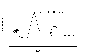
WHAT IS SCATTER PLOTS?
Laser light is shown on the cell. Light is scattered in 4
different directions.
The scattered light is analysed by the instrument.
The WBC features like Granularity, Lobularity (number of
lobules in nucleus) Size of the cells can be identified
Scatter plot shows - as dots the distribution of each cell
which the machine has seen
HAEMOGRAM
RBC Histogram shows
Size in X axis
Number in Y axis
Average size of an RBC is 7.5 micron in diameter (Recticulocyte
9µ)
Number 4 – 6 million / cu.mm
The relation between the size and number is reported as RDW
in computer print out.
WHAT IS RDW ?
The width of RBC peak in the histogram is called the Red
cell Distribution Width (RDW). It shows the heterogenecity of RBC population-
more variation in size is expressed as large RDW. This is mentioned as
anisocytosis and poikilocytosis by pathologist on seeing peripheral Smear.
Normal range of RDW – 11.5 - 14.5%
RBC Count = Normal value - 5 Million / cu.mm
Hb Normal value is 15 gm/dl
PCV is % of RBC in total blood volume. HCT means PCV. Normal value is 45 %
WHAT IS RULE OF 3 for a normal person (not for disease
states)
RBC count in million x 3 = Hb in gm/dl
Hb in gm/dl x 3 = PCV
Eg. RBC count, 5 m/cu..mm x 3 = 15 gms/dl of Hb
15 gms/dl x 3 = 45% PCV
This relationship is disturbed in disease states. So if you
find such an abnormality in the result, it indicates an underlined disease in
the patient which can be diagnosed precisely if all these data are correlated.
RBC indices are
MCV = Average volume of an RBC
MCH = Amount of Hb in one RBC
MCHC = % of Hb in RBC
MCV varies with age. Normal values are given in
Table 1
MCH normal value is 29 – 30 Picogram
MCHC normal value is 33 – 34 gd/dl
HAEMOGRAM
Low MCV = Microcyte
Normal MCV = Normocyte
High MCV = Macrocyte
Low MCH = Hypochromic
Normal MCH = Normochromic
Reticulocyte count is not directly done by this machine
but, it is mentioned as NRBC. So a raised NRBC means Reticulocytosis.
A few examples of Abnormal result (i.e., the rule of 3 not
observed) is:
Cold Agglutinin (Paroxysmal Cold Haemoglobinuria)
Hemolysis States (Incompatible Blood Transfusion, Burns,
Neonatal Jaundice, Prosthetic Valve, etc.)
Hyper lipidema
Hyperglycemia
Micro clots (Prosthetic Valve, Infective Endocarditis,
etc.,)
blood transfusion
Abnormal values can be due to instrument fault or disease
states. If instrument calibration is normal then disease state should be
suspected. For detecting this disease state a clinical data with the
requisition for Haemogram is essential. If there a machine error, the print
out does not come out.
BENEFIT FOR PATIENTS
- cost less
- quick results
- waiting period to get result is less
- only small amount of blood needed. So repeated sampling is not difficult
eg. In cancer patients. In
- newborns small amount only may be obtained. < 1 ml will be sufficient
if collected Properly.
BENEFIT FOR DOCTORS
- Quick result
- Interpretation helps in reducing number of investigation ordered.
- Eg. Anemia work up is very easy
- No need to wait for Pathologists report
- In many cases peripheral smear exam may not be needed
A few examples of how haemogram helps you to come to definite diagnosis is
illustrated here.
-
52 year old man with fever of 1 week duration.
The haemogram shows
WBC Count 3200 cells/cu.mm
Neu 12%
Lym 80%
Mono 4%
Hb 7 gm/dl
Platelet count 40,000/cu.mm. Machine interprets results are Leukopenia,
Neutropenia, Anemia, Thrombocytopenia.
Here the diagnosis is Aplastic anemia because RBC,WBC, and Platelet conts
are all decreased.
This could have been diagnosed conventionally by doing all the above
parameters manually and doing peripheral smear with Pathologists’s report.
-
8 year old child with fever and breathlessness of 2 days duration.
Haemoram shows
WBC count 18,000 cells /cu.mm
Neu 80%
Lymph 12%
Mono 8%
RBC normal values
Platelets count 420,000 /cu.mm
Machine interprets results as Leukoytosis, Neutrophilia, Monocytosis,
Thrombocytosis
Immature Granulocytosis.
These values indicate acute bacterial infection – may be pneumonia if
related with clinical details.
A COMPARISON BETWEEN PERIPHERAL SMEAR & HAEMOGRAM
|
Disease |
Peripheral Smear Results |
Haemogram Results |
Investigations to confirm Diagnosis |
|
Iron Deficiency Anemia |
small RBC |
low Hb, low MCV, low MCH, and low MCHC, high RDW |
Serum Iron, TIBC, Ferritin, Bone Marrow staining with Prussian Blue |
|
Megaloblastic Anemia |
large RBC |
Low Hb, high MCV, high RDW
|
B-12, Folic Acid deficiency to be investigated such as Nerve conduction
study, FIGLU |
|
Thalassemias |
small RBC, target cells |
low MCV, low MCH, low MCHC, high RDW |
HbF, Osmotic Fragility, Hb Electrophoresis |
|
Haemolytic – Uremic Syndrome, Burns |
Schistocytosis (Anisocytosis) (Fragmented RBCs) – Burr cells, Helmet
cells |
high RBCs & Platelet counts,
low MCH & MCHC |
Blood Urea, and other Renal function studies |
|
Paroxysmal Cold Haemoglobinuria |
Rouleux formation |
low RBC count
two population of RBC Histogram |
Repeat Haemogram with warm blood will give normal Haemogram |
|
Blood transfusion |
No specific findings |
2 population of RBC one of patient & one of donor |
|
[top]
|